
More Information
Submitted: May 10, 2024 | Approved: May 25, 2024 | Published: May 27, 2024
How to cite this article: Ali MN, Hasan M, Shanta IS, Choudhury MA, Rahman M, et al. Myiasis in a Backyard Pig: A Case Report. Insights Vet Sci. 2024; 8: 015-017.
DOI: 10.29328/journal.ivs.1001042
Copyright License: © 2024 Ali MN, et al. This is an open access article distributed under the Creative Commons Attribution License, which permits unrestricted use, distribution, and reproduction in any medium, provided the original work is properly cited.
Keywords: Myiasis; Backyard pig; Clinical management

Myiasis in a Backyard Pig: A Case Report
Md Niamot Ali1, Mahdi Hasan2, Ireen Sultana Shanta2, Md Abu Choudhury3, Mustafizur Rahman2 and Abdulla Al Mamun Bhuyan1*
1Department of Veterinary & Animal Sciences, University of Rajshahi, Rajshahi-6205, Bangladesh
2International Centre for Diarrhoeal Disease Research, Bangladesh, Mohakhali, Dhaka-1212, Bangladesh
3Department of Nursing and Allied Health, Swinburne University of Technology, Melbourne, Australia
*Address for Correspondence: Dr. Abdulla Al Mamun Bhuyan, Department of Veterinary & Animal Sciences, University of Rajshahi, Rajshahi-6205, Bangladesh, Email: bhuyan@ru.ac.bd
Background: Myiasis is a parasitic infestation of livestock animals caused by dipteran larvae. The presence of wounds, lack of hygiene on the farm, and temperate climatic conditions contribute to myiasis. Swine can be infested by myiasis if injured pigs are not treated properly and failure to treat myiasis in time may cause the culling or death of the pigs, resulting in huge economic loss to the farmers. But like humans and other farm animals, pigs also deserve to be treated and cured of any suffering or disease. Therefore, this study is documented on pig myiasis and its management because to date a few cases have been reported on it.
Case presentation: This case report documented the successful management of neck myiasis in a male, 9-month-old, 12-kg-weighing backyard pig. The wound site was cleaned using antiseptics and maggots were removed. The site was treated with turpentine oil, and ivermectin at 0.2 mg/kg B.W. and S/C. A combination of streptomycin (12.5 mg/kg B.W.) and penicillin (20000 IU/kg B.W.) was used IM daily for 5 days to prevent secondary bacterial infection. The wound was dressed regularly on every alternate day until the complete removal of maggots and the formation of granulation tissue.
Conclusion: Through proper therapeutic management, the backyard pig’s neck myiasis wound was successfully healed in 10 days without any complications.
Pigs are highly fertile animals and well-known for their large litter sizes [1]. In comparison to other livestock, they are two times more efficient at converting food into meat [2,3]. Genetically, pigs are high meat producers, and the average ratio of feed conversion is around 3.00 [4,5]. Pigs are found and raised around the world, and they provide valuable products to human beings, including meat, lard, hide, adhesives, fertilizers, and various pharmaceuticals. According to the USDA report (2022-23), most of the pigs (78% of total pigs) in the world are raised in China, the EU, and the United States, and they roughly contribute 48%, 19% and 11% of total pork production, respectively [6]. In the United States, they produce more lean meat than lard, a fat used in cooking [7]. Pig meat is considered the richest but comparatively cheaper protein source [8]. According to the FAO report (2013), the significant pork-consuming countries in the world are Austria, Germany, Spain, Poland, Italy, Netherlands, and France, and they consume 51-52 kg/capita/ year, 51-52 kg/capita/ year, 49 kg/capita/ year, 46 kg/capita/ year, 40kg/capita/ year, 36 kg/capita/ year, and 33kg/capita/ year respectively [9].
Due to religious beliefs and taboos, only non-Muslim minorities like Christians, lower-class Hindus, Sweepers, ethnic or tribal peoples, the Cobbler community, and nomadic peoples raise pigs in Bangladesh [10-12]. As a result, most of the people in Bangladesh are not familiar with pig farming. Currently, pig farmers are not aware of the treatment of injured or diseased pigs due to their illiteracy and communication gap with the veterinary services in Bangladesh [13]. Like other livestock, pigs also suffer from many diseases, including classical swine fever, foot and mouth diseases (FMD), brucellosis, anemia, injury, mange, myiasis, etc.
Adult pigs are most susceptible to injury or wounds, followed by myiasis if untreated [14,15]. Myiasis is an external parasitic infestation caused by fly larvae in live animals or humans [14,15]. Unhygienic conditions and tropical weather favor the prevalence of myiasis [16]. Female flies lay eggs [17] at the wound area when feeding the oozing. Fly eggs are usually licked off wherever the animal can reach their tongue at the site of a wound. There are three different larval stages of fly (1st Instar, 2nd Instar, and 3rd Instar), depending on the time of infestation and larval development [17]. Larvae in tissue cavities cause unwanted inflammatory reactions, bacterial infections, or granuloma formation [18]. Areas at risk for animals where larvae frequently infest are the hind limb, ear, fore limb, back of the body, shoulder, tail, and vulva [19]. Although myiasis is relatively common in cattle and goats (46.4%), pigs also suffer (6%) [16]. The surgical affections of the pigs are considered a threat to their productive capabilities, market values, and welfare. Immediate care against myiasis relieves the suffering of the pigs and saves production losses. Failure to treat may result in the culling or death of the pigs. The study aims to heal the myiasis wounds of the pigs and improve animal welfare by decreasing animal suffering.
The Research titled ‘Study design and operation for Nipah virus vaccine candidate among backyard pigs of the indigenous communities in Rajshahi, Bangladesh’ was conducted in a pig-raising community in Rajshahi, Bangladesh. During this study time, a pig owner complained about a backyard pig having a wound in its neck region for the last few days with the following identification: Sex: male; age: 9 months; body weight: 12 kg; and coat color: black. The owner also reported scratching the neck against any hard objects. Our investigation revealed the pig was tightly secured on its neck with a nylon rope, causing an incised wound. Close inspection and physical examination found the presence of numerous larvae in the wound (Figure 1), confirming the case of myiasis.
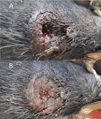
Figure 1: Neck Myiasis in a Backyard Pig.
After proper restraining of the pig, the nylon rope was removed, and the wound and its surrounding area were cleaned thoroughly by removing all of the tissue debris and dirt with an antiseptic solution. Then, superficial maggots (1st and 2nd instar stage of larva) were removed using forceps. Oil of turpentine was applied locally, and dead maggots were removed the following day. The pig was administered to Inj. Ivermectin at 0.2 mg/kg B.W. (Inj. Vermic Vet 5ml; Techno Drugs Ltd. at S/C route). As post-operative care and to prevent further complications from secondary bacterial infection, systemic antibiotics like a combination of penicillin at 20000 IU/kg B.W. and streptomycin at 12.5 mg/kg B.W. (Inj. SP-Vet 2.5 g; ACME Laboratories Ltd.) were given IM daily for 5 days. Dressing of the wound was done with Tr. of iodine as an antiseptic wash every other day until the granulation of tissue started to grow. The pig was kept in a confined area instead of tying it up with rope. The owner was suggested to spray turpentine oil 4-6 times a day around the wound to prevent further fly infestation. The wound healed in 10 days without any further complications.
Myiasis is a parasitic infestation of living tissues with dipteran fly larvae. Larvae ingest the tissue, tissue fluid, and tissue debris of the wound. There are four different types of myiasis reported: bloodsucking myiasis, cutaneous myiasis, wound myiasis, and cavitary myiasis [15,20]. Adult male pigs are less susceptible to myiasis than females, but young male pigs are more affected than adults [19]. The victim of this case is also a young pig affected by both wounds and cavitary myiasis. The incidence of myiasis is more frequent from June to November [19,21], but it has the lowest incidence from December to February in southeast Asia [19]. The present case was also reported in October, which indicates the influence of the season on the occurrence of myiasis. Burgess, in his study, also reported that myiasis is relatively more frequent and common in tropical regions [22]. In dairy animals, myiasis is the most frequent, and most incidences are observed around the hooves [23]. In pigs, myiasis at the hind limb is most prevalent, but neck myiasis is also reported in higher numbers [19]. Myiasis in this case report was found in the necks of the pigs. Tightly securing the pig with the nylon thread through the neck works as a predisposing factor in creating the wound. Oozing from the wound attracts the fly to infest it. Wound healing depends on various factors, including aging, medications, obesity, blood circulation, and the presence of infection in the wound [24]. Healing may be delayed when wounds are infected by microorganisms, swollen or edematous, and contain any foreign bodies or necrotic tissues [25]. Thorough irrigation of the wound and dressing is necessary to improve the healing process and save the animal from further complications like abscess, gangrene, or septicemia and death [26,27].
The backyard pig’s neck myiasis wound was successfully healed in 10 days without any complications through proper therapeutic management.
Authors’ contributions
Mahdi Hasan and Abdulla Al Mamun Bhuyan carried out the treatment of the pig. Md. Niamot Ali developed the draft of the report. Ireen Sultana Shanta, Md Abu Choudhury, Mustafizur Rahman, and Abdulla Al Mamun Bhuyan revised the manuscript. All authors reviewed and approved the final manuscript.
The research group is thankful to the Department of Veterinary and Animal Sciences, University of Rajshahi, Bangladesh, for facilitating the treatment and research, and the International Centre for Diarrhoeal Disease Research, Bangladesh, Mohakhali, Dhaka-1212, Bangladesh, for funding the project.
- Njoga UJ, Ilo SU, Ugwu PC, Ajibo FE, Bernard NS, Onwuka OS, Njoga EO. Reproductive and fertility parameters of pigs reared in Enugu State, Nigeria, Animal Research International. 2021; 5;18(1):3918-26.
- Mpofu I, Makuza SMM. Pig Production Science and Technology, 1st edition, Ed: A. Shonhiwa, Upfront Publishing, UK. 2003.
- "Germany: Better performance and increased litter size - Pig Progress" https://www.pigprogress.net/health-nutrition/germany-better-performance-and-increased-litter-size.
- Losinger WC. Feed-conversion ratio of finisher pigs in the USA. Prev Vet Med. 1998 Oct 9;36(4):287-305. doi: 10.1016/s0167-5877(98)00094-4. PMID: 9820889.
- Wang D, Huang J, Lohmar B. Feed Conversion Ratio, Profitability and Farm Size in China’s Pig Industry, 2015.
- Pork | USDA Foreign Agricultural Service. USDA Foreign Agricultural Service.
- "Pig" https://kids.nationalgeographic.com/animals/mammals/facts/pig.
- Feed-to-Meat - Conversion Inefficiency Ratios - A Well-Fed World. https://awellfedworld.org/feed-ratios.
- Szűcs I, Vida V. Global tendencies in pork meat-production, trade and consumption. Appl Stud Agribus Commer. 2017 Dec 31;11(3-4):105-11.
- Anower AK, Ahmed M, Rahman MM, Hasan A, Islam MA, Rahman L. Hygienic farming system improved pig-rearers livelihood status in South-West region of Bangladesh. Int J Avian Wildl Biol. 2017; 2:91-97.
- Khanum R, Mahadi MSA, Islam MS. Tribal women's involvement with pig farming in Bangladesh: an evidence of Moulvibazar district. SAARC J Agric. 2018; 16(1):115-127.
- Bangladesh Bureau of Statistics. Report of the household-based livestock and poultry survey 2009. In: Division S, editor: Ministry of Planning, Government of the People’s Republic of Bangladesh; 2010.
- Islam A, Trisha AA, Safiul M, Sardar A, Akbor M, Al Mamun A, Bhuyan MS, Faruk MA, Sharif SM, Nahar Z. Pig raising practices by underprivileged, ethnic people in Bangladesh. Insights Vet Sci. 2021; 15(25):35.
- Zumpt F. Myiasis in man and animals in the Old World, Butterworths, London, United Kingdom. 1965.
- Francesconi F, Lupi O. Myiasis. Clin Microbiol Rev. 2012 Jan;25(1):79-105. doi: 10.1128/CMR.00010-11. PMID: 22232372; PMCID: PMC3255963.
- Imtiaz MA, Rahman MA, Islam K, Barua M, Alim MA, Chowdhury S, Sikder S. Prevalence and associated risk factors of myiasis in different areas of Chittagong, Bangladesh. Res J Vet Pract. 2014;2(2):22-7.
- Glory M, Paul N. Study on the development of maggots on meat, International Journal of Science and Research Achieve, 2024.
- Sunny B, Sulthana L, James A, Sivakumar T. Maggot Infestation: Various Treatment Modalities. J Am Coll Clin Wound Spec. 2018 Mar 30;8(1-3):51-53. doi: 10.1016/j.jccw.2018.03.002. PMID: 30276127; PMCID: PMC6161638.
- Chakrabarti, ASIT. Incidence of maggot wound in crossbred pig in an organized farm. In The International Conference on Integrating Climate, Crop, Ecology-The Emerging areas of Agriculture, Horticulture, Livestock, Fishery, Forestry, Biodiversity and Policy Issues at Jawaharlal Nehru University, New Delhi, 4th June. 2016.
- Veraldi S, Brusasco A, Süss L. Cutaneous myiasis caused by larvae of Cordylobia anthropophaga (Blanchard). Int J Dermatol. 1993 Mar;32(3):184-7. doi: 10.1111/j.1365-4362.1993.tb02789.x. PMID: 8444529.
- Chakrabarti A, Kumar D. Disease Incidences in Pigs due to Seasonal Variation and Climatic Effect in an Organized Farm, 2017.
- Burgess IF. Myiasis: maggot infestation. Nurs Times. 2003 Apr 1-7;99(13):51-3. PMID: 12715561.
- Singh A, Singh D. A study on the incidence of myiasis among dairy animals in the State of Punjab, India. 2016; 9(1): 30-34.
- Grada A, Mervis J, Falanga V. Research Techniques Made Simple: Animal Models of Wound Healing. J Invest Dermatol. 2018 Oct;138(10):2095-2105.e1. doi: 10.1016/j.jid.2018.08.005. PMID: 30244718.
- Kamble S, Ganguly S, Qadri K, Mahajan T. Management of Auricular Myiasis in Swine: A Case Report. Int J Contemp Pathol. 2016; 2(1):49-50.
- John, D. and Petri, W. Markell and Voge’s Medical Parasitology, 9th Ed, Missouri: Saunders Elsevier. 2006; 328-334.
- Jervis-Bardy J, Fitzpatrick N, Masood A, Crossland G, Patel H. Myiasis of the ear: a review with entomological aspects for the otolaryngologist. Ann Otol Rhinol Laryngol. 2015 May;124(5):345-50. doi: 10.1177/0003489414557021. Epub 2014 Oct 30. PMID: 25358614.